Postnatal Heart Development & In Vivo Functional Genomics
We define the cellular, molecular, spatial, and functional programs that guide cardiomyocytes from postnatal growth toward mature heart function.
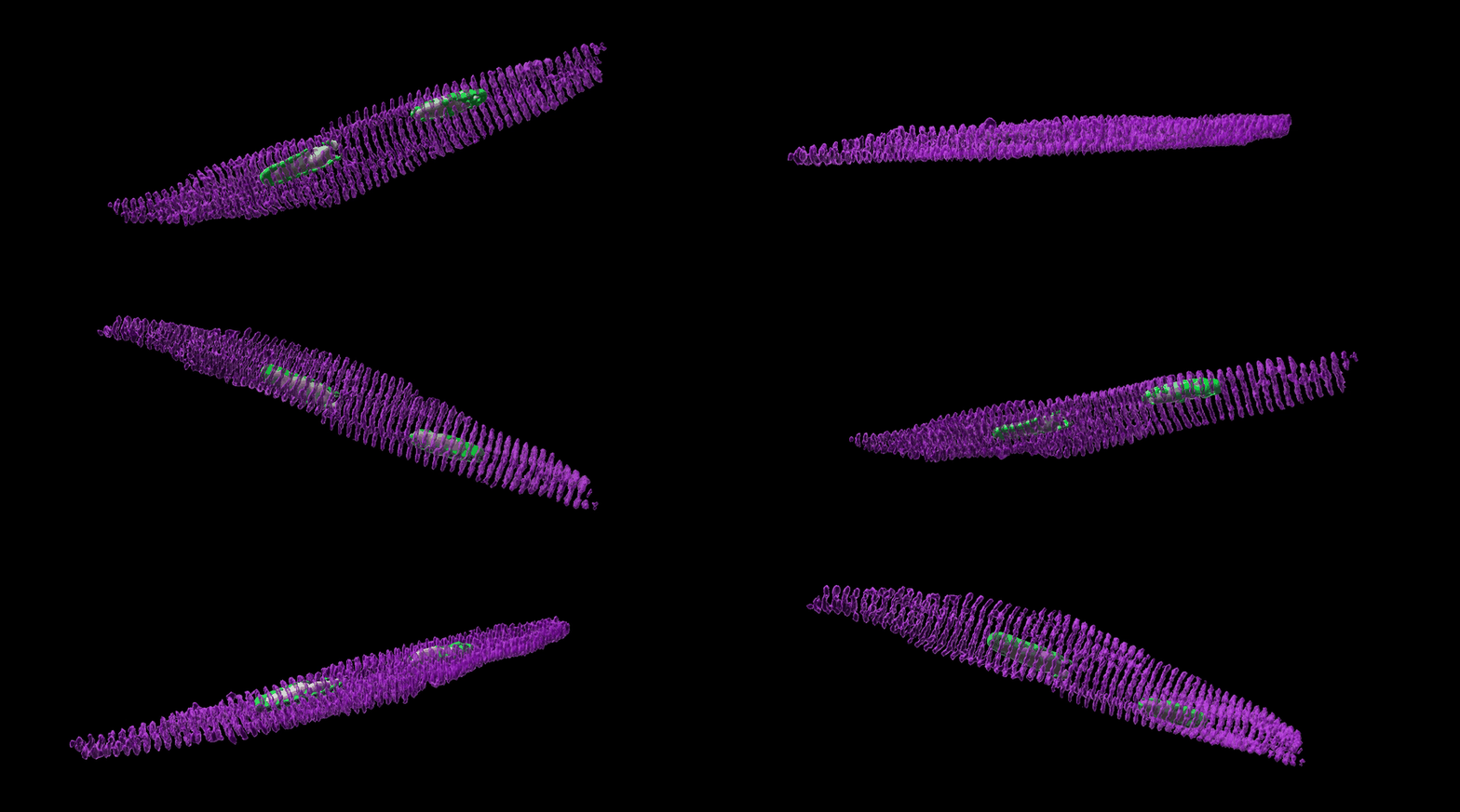
§ Understanding How the Heart Becomes Mature
The mammalian heart undergoes a remarkable transformation after birth. During this period, cardiomyocytes transition from relatively immature, proliferative cells into highly specialized muscle cells capable of sustaining billions of contractions over a lifetime. This maturation process involves coordinated changes in cell size, metabolism, contractile organization, electrical activity, gene regulation, and communication with neighboring cell types.
Despite its importance, we still do not fully understand how these developmental programs are coordinated or which molecular regulators are required for cardiomyocytes to acquire and maintain a mature state.
Our research seeks to define the cellular, molecular, and spatial mechanisms that drive postnatal heart maturation.
§ Why Study Heart Maturation?
Many forms of heart disease involve the loss of maturity and the gain of fetal-like features of cardiomyocytes. Additionally, cardiomyocytes generated from pluripotent stem cells often remain developmentally immature, limiting their usefulness for disease modeling, drug testing, and regenerative therapies.
A deeper understanding of normal heart maturation can help us:
- Identify molecular pathways that promote mature cardiomyocyte function
- Improve the maturation of stem-cell-derived cardiac cells
- Reveal why the adult heart has limited regenerative capacity
- Discover developmental programs that become disrupted in heart disease
- Develop new strategies to repair damaged myocardium
Rather than viewing maturation as a single developmental endpoint, we study it as a coordinated process shaped by gene-regulatory networks, cell-cell interactions, tissue architecture, and changing physiological demands after birth.
§ Mapping the Postnatal Heart
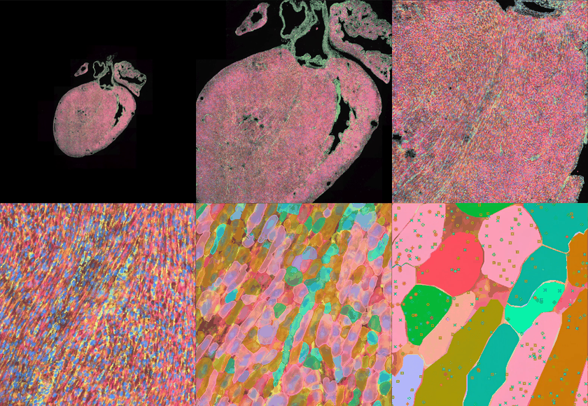
The heart is composed of diverse cell types organized into distinct anatomical and functional environments. Cardiomyocytes mature within these local tissue niches while interacting with endothelial cells, fibroblasts, immune cells, and other neighboring populations.
We combine single-nucleus RNA sequencing, spatial transcriptomics and computational analysis to construct detailed maps of the developing postnatal heart. These approaches allow us to examine:
- How cardiomyocyte states change over developmental time
- How maturation differs across ventricular regions
- How cardiac cell populations are spatially organized
- How local cellular environments influence cardiomyocyte development
- How communication between cardiomyocytes and non-myocytes changes after birth
By integrating molecular and spatial information, we aim to understand not only which genes are expressed during maturation, but also where, when, and in which cellular context these programs operate.
§ Moving from Correlation to Function
Genomic and spatial profiling can identify thousands of genes associated with heart development. However, association alone does not establish whether a gene is required for maturation or whether changing its activity can improve cardiac function.
To address this challenge, we develop and apply in vivo Probe-based indel-detectable Perturb-seq (PIP-seq) to test candidate regulators directly in the postnatal heart.
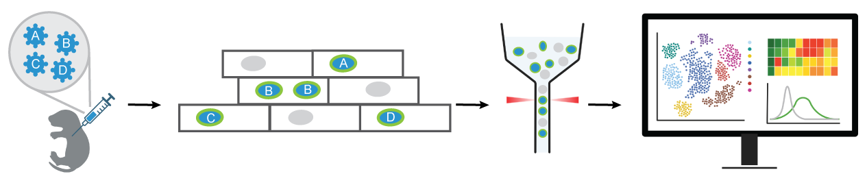
Using in vivo PIP-seq, we can evaluate many candidate genes within their native physiological environment in high-throughput. The whole transcriptome readout allow us to determine how individual regulators affect cardiomyocyte growth, metabolism, contractile organization, electrophysiology, and gene expression.
§ Why In Vivo Functional Genomics?
Cardiomyocyte maturation is strongly influenced by the environment of the living heart. Mechanical force, blood flow, oxygen availability, circulating hormones, electrical activity, extracellular matrix, and signals from neighboring cells all contribute to the maturation process.
These conditions are difficult to fully reproduce in conventional cell culture.
In vivo functional genomics allows us to study gene function while preserving: Native cardiac tissue architecture; Cell-cell and cell-matrix interactions; Normal vascular and metabolic environments; Systemic endocrine and neuronal signals.
Our goal is to bridge the gap between large-scale genomic discovery and physiological function. By combining spatial mapping with in vivo perturbation, we can prioritize regulators based not only on where they are expressed, but also on what they actually do in the developing heart.
§ Long-Term Vision
Our long-term goal is to uncover the regulatory principles that allow cardiomyocytes to achieve mature function while maintaining the health and stability of the heart.
By understanding how maturation is controlled in vivo, we hope to establish a foundation for:
- Producing more mature stem-cell-derived cardiomyocytes
- Building more accurate human cardiac disease models
- Identifying therapeutic targets for developmental and acquired heart disease
- Improving cardiac regeneration and cell-based therapies
- Restoring mature cardiac function after injury
Ultimately, we aim to transform developmental knowledge into new strategies for understanding, modeling, and treating heart disease.